


Lumbar osteochondrosis is a dangerous disease of the spine, characteristic of people who have reached the age of 35 years and older. The natural wear and tear of the joints causes the development of pathology. Improper access to rheumatologists, in most cases, leads to disability. Modern medicine offers many effective treatment methods at a major stage. Early diagnosis is the key to a healthy life without restrictions.
Lumbar osteochondrosis - a general definition
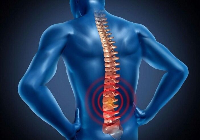
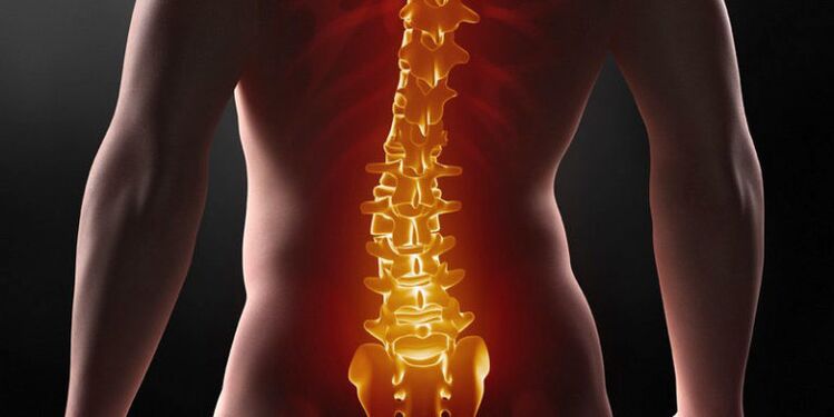
Osteochondrosis of the lumbar spine is a process of dystrophic degeneration in the formation of intervertebral cartilage - discs.
Discs provide the main functions of the spine - the ability to move and bend, resistance to stress. As a result of pathology, vital elements become thinner, deformed, vertebrae are aligned, nerve endings and blood vessels are pinched. The negative process is accompanied by pain sensations of varying intensity and limits of motor function.
Pathology causes changes in the connecting elements of the spine - cartilage, bones, discs and joints. It is caused both by natural wear and tear processes, and by diseases acquired in the joints or as a result of an improper lifestyle.
cause
There are many reasons for the development of lumbar osteochondrosis:
- Natural or premature wear of the body;
- Excessive load on the lower back - lifting weights, working "on your feet" or an inactive, "sedentary" lifestyle;
- Genetic predisposition to joint diseases, such as rheumatoid arthritis;
- Violation of metabolism, resulting in accumulation of toxic substances in the connecting disc;
- Chronic diseases of the circulatory system. Nutrients and trace elements stop entering cartilage tissue in proper amounts. Hypoxia occurs, which contributes to the destruction of the intervertebral joints;
- Autoimmune pathology.
Secondary factors can also trigger the development of lumbar osteochondrosis:
- Chronic injuries, back bruises;
- Exceeding the weight norm by more than 15-20%;
- Heavy or powerful sports;
- Always wear uncomfortable shoes. High heels, tight shoes, rubber or sports shoes are the first enemies of the spine;
- Valgus changes in the legs;
- Scoliosis, kyphosis, diabetes mellitus, spinal tuberculosis;
- Low temperature effect.
Clinical picture
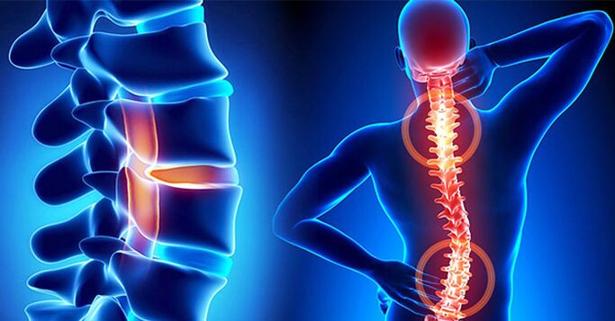
The symptoms of lumbar osteochondrosis depend entirely on which nerve root is affected by the disease. The degree of vertebral compression, the stage of the disease and damage to the disc determine the symptoms.
Rheumatology distinguishes the following main symptoms:
- Violation of tactile susceptibility in the lumbar region. Numbness extends to the inside of the thighs and groin. May affect one or both limbs;
- There is a sharp pain in the lower back. The big toe completely loses mobility and characteristic numbness is observed;
- Loss of normal function of the legs, sensitivity of the fingers, lower legs and outer thighs. In this part of the foot, there are frequent tones and seizures. On examination, there was no Achilles reflex;
- If the disease affects the lower radicular arteries, then there is complete paralysis of the muscles of the back, back of the thighs and perineum. There is a serious violation of motor function, so that it can not move fully.
With lumbar osteochondrosis, not only the nerve endings of the spine are affected, but also the blood vessels.
The following specific signs depend on the type of lesion:
- When only the nerve roots are disturbed, changes in the patient’s gait are observed. The pain is localized not only in the lumbar zone, but also in all parts of the leg. Radicular syndrome is characterized by persistent pain. Usually just next door. In the lower back, tingling and pain are observed. The pain can be relieved with a little exercise.
- Compression of the blood vessels leads to perfusion in the hip area. As a result, oxygen starvation of the spinal disc occurs. Painful sensations occur while walking in the buttocks, thighs and lower back. Completely discarded after an overnight rest.
Simultaneous violation of the function of blood vessels and nerve roots can lead to irreversible disc deformation. Spiny -shaped bone plants form in the movable joints of the lower back. This leads to severe pain and makes normal natural movement impossible. Violated posture, walking style. When it does occur, complete paralysis may occur.
Disease stage
Lumbar osteochondrosis develops gradually, in several stages. Each level has its own characteristics, which determine the level of progress.
- I'm on stage.Slow destruction of the intervertebral disc begins. This process can last from a few months to 2-5 years. Manifested by minor pain, discomfort in the inguinal and femoral muscles. It is observed while walking or when the weather changes.
- stage II.The collagen fibers of the spinal fibrous ring are pulled into a negative process. The space between individual vertebrae shrinks rapidly. Friction appears, which causes a severe attack of pain. Violated walking style, posture, crouching appear. Lumbar osteochondrosis is most often diagnosed in the second stage of the course.
- Level III.An intervertebral hernia appears. And if the patient is not forced to seek medical help with stage II symptoms, then it is no longer possible to ignore the excruciating pain in the third stage. The deformity of the bones and joints of the spine in the lumbar zone is no longer irreversible. Walking takes a lot of effort. This is due to the pain and the inability to relieve it with conventional painkillers.
- stage IV.Partial or complete deterioration of motor function. At this stage, patients are assigned a disability group. Threatens with complete paralysis. Vital activities are impossible without taking various types of medications.
Diagnostic measures
Diagnostic measures include several techniques and begin with the collection of a complete history of the disease. During the initial consultation with a rheumatologist, the following data are explained:
- Patient complaints are carefully analyzed - the place of localization of pain, where discomfort is still felt, where the part of the hip joint there is a feeling of heaviness, seizures, etc. ;
- Duration, regularity, nature of pain;
- When the first symptoms, even minor, appear. How long has passed since the last attack, what caused the discomfort and what factors contributed to its elimination;
- Living conditions around the patient. Profession, work, household load, sports and the presence of additional factors for increased physical activity (dacha, garden, hobbies related to weight transfer);
- Examination of the history of disease that the patient has had before or now.
After collecting the clinical picture, the rheumatologist proceeded directly to the external examination. During the examination, gait, anatomical position of the legs, arms, body, in relation to the spine were analyzed. The skin is examined for changes - pigmentation, exfoliation, eczema, rashes, etc. Assessment of motor function is given.
Performing simple exercises - leaning forward, backward, lifting arms and legs, turning the head, rotational movements of the pelvis, the patient allows the doctor to determine the degree of damage to the spine in the lumbar region.
The final step of external inspection is the action to determine the degree of radicular damage:
- Lasegue symptoms.Lying on his back, the patient lifts the legs alternately, bent at the knees. If this causes pain in the lower back, then the reading is considered positive.
- Symptoms of Dejerine.The patient is asked to tighten the abdominal muscles as much as possible. The occurrence of discomfort in the spine indicates the development of lumbar osteochondrosis.
- Symptoms of Neri. A sharp tilt of the head forward and backward responds with pain in the lower back.
- Wasserman's symptoms. The patient, in the supine position, moves the leg to the side as much as possible. In the presence of pathology, unpleasant pain occurs in the groin and front of the thigh.
To confirm or exclude a diagnosis, patients are invited to undergo an instrumental diagnosis. MRI is considered the most effective way to determine lumbar osteochondrosis. Studies show the distance between the vertebrae, the development of neoplasms and bone defects. It may be contraindicated in patients with mental disorders.
Computed tomography gives a fairly true picture of the disease in one plane - horizontal or vertical.
X-rays are used only in the last stage, when irreversible changes in the spinal bone tissue begin.
Complex treatment of lumbar osteochondrosis
The pathological causes have not been fully elucidated. Scientific research in the field of spinal articular diseases has not identified a sufficiently effective method for complete rehabilitation of the intervertebral disc. Modern treatment methods are only aimed at eliminating the outward signs of the disease. Full recovery is currently considered impossible.
Traditional medicine therapy
Rheumatologists prescribe medication, depending on the general condition of the patient. The clinical picture provides the information needed to create a treatment regimen with drugs from several groups.
- Anesthetic agent.Injections, ointments or broad -spectrum medications are prescribed.
- Anti-inflammatory drugs (NSAIDs).
- Vasodilator.Removal of tone from the lumbar and leg muscles.
- Chondroprotectors.Designed to exclude negative progression of lumbar osteochondrosis.
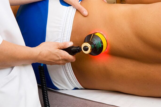
Physiotherapy
Physiotherapy procedures are part of the inpatient or outpatient treatment of lumbar osteochondrosis.
Includes the following activities:
- Electrophoresis with painkillers;
- Magnetotherapy;
- Hydrotherapy;
- Paraffin application.
Medicine and physiotherapy in the complex relieve pain and acute inflammation. But they are not a guarantee to stop the pathological progression. Only a course of treatment 2-3 times a year and a responsible attitude of the patient will help to avoid regression and maintain the general condition in a satisfactory shape.
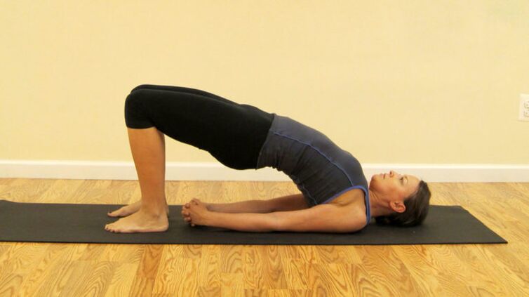
Exercise therapy and therapeutic massage
A set of therapeutic gymnastic exercises ensures the normalization of blood circulation in the lower back and helps eliminate the process of stagnation. Only a physical therapist can prescribe exercises for clinical or home use. As a rule, these are all kinds of gentle tendencies and rotational movements, from prone and sitting positions. Independent physical activity not only does not bring results, but causes more displacement of the vertebral discs.
Manual therapy sessions help strengthen muscle tissue, blood flow to the affected lower back, and relieve tension. Specialists perform a massage first on a healthy back, to warm up the muscles and improve blood circulation. Then it goes to the affected lumbar area. Areas of manipulation include the lower back, buttocks, thighs, hamstrings and legs. Sessions are held in regular courses, at least 10 sessions in 6 months.
Surgical intervention
It is indicated in the last stages of lumbar osteochondrosis, to restore spinal motor function. Surgery remains the only option for patients with the following symptoms:
- Persistent pain syndrome, unable to receive treatment even with opiate -containing medications;
- Strong compression of nerve roots and significant displacement of the disc;
- Neoplasms, proliferation of bone tissue;
- Complete destruction of the vertebrae, due to constant friction;
- Paralyzed.
Modern methods offer less traumatic methods of internal intervention. For example, endoscopy. It has a favorable prognosis, a short recovery period and a low rate of side effects.
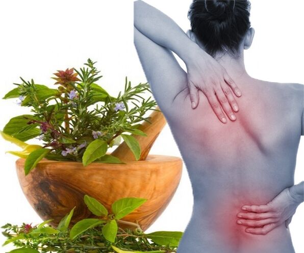
Alternative treatments
Lumbar osteochondrosis responds well to treatment with medicinal herbs and folk methods. Ointments, tinctures, baths based on fees are used to relieve swelling and pain. The most effective recipes include anesthetic and anti-inflammatory herbs:
- yarrow;
- Aloe;
- Mint;
- St. WortJohn;
- Spruce or pine needles;
- Sage.
The content of these herbs in folk recipes is due to their medicinal effects, scientifically proven by traditional medicine. Home treatment will help keep the lower back in a stable condition and prevent worsening of the disease after complex treatments.
Prevention
Despite the fact that lumbar osteochondrosis is an incurable disease, its negative manifestations can be minimized. In the early stages, the disease is successfully treated, only need to seek timely medical help. It is important to fully adhere to the treatment regimen provided and follow the recommendations of the rheumatologist.